for students

Bachelor and Master students
We always welcome Bachelor and Master students in our lab. Please see below for available projects.
Bachelor students can participate in a ten-week lab course called "Forschungspraktikum II mit Bachelorarbeit" in our lab during their third year. Applications for this lab course are due in the fall semester for the following spring.
If you are interested in becoming a Master student in our lab, you typically apply during the spring semester. However, alternative application dates can be arranged.
We also offer summer lab courses as a substitute for one of the mandatory "Semesterarbeit" (6.5 ECTS). The application deadline for this lab course is in late October for the following summer break.
If you are simply curious to see what it's like to work in a lab, you can join us for some time during the summer.
Currently available Master projects
(i) EXPANDER: Expansion microscopy of protein-complex arrangement in the mitochondrial membrane
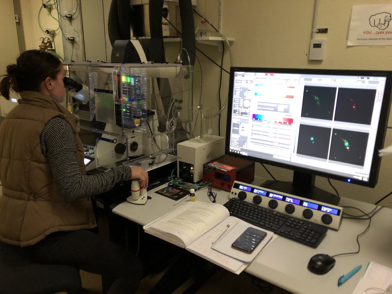
Expansion microscopy is a relatively new approach that has been added to the super-resolution microscopy toolbox and is expected to have a significant impact on the field of microscopy. This technique uses expandable hydrogels, similar to those found in diapers, to increase the physical distance between two components of interest in the cell isotropically, resulting in an increase in resolution of up to five- or even tenfold. In simple terms, it is like blowing up the cell like a balloon. This technique will enable us to address various questions related to the positioning, interaction, and biogenesis of proteins/complexes in the cell. Our project aims to develop the technique further to elucidate the precise architecture of the mitochondrial genome segregation machinery in the mitochondrial membranes. Previous studies have shown the architecture of the complex at a lower resolution, and we anticipate being able to achieve a resolution below five nanometers using this technique in combination with STED super-resolution microscopy.
Impact:
• Expansion microscopy is a cutting-edge technology for localizing proteins, and our lab is the first to use it in trypanosomes. • The positioning of proteins in the outer mitochondrial membrane relative to the inner mitochondrial membrane is currently challenging to observe using fluorescence microscopy due to the short distance between the two membranes. • However, expansion microscopy is a game changer for studying complex interactions because it allows us to increase the distance between these membranes and visualize the proteins with greater resolution.
What will you do:
In this project the student will be trained to prepare expansion microscopy samples and then characterize positioning of several mitochondrial membrane complexes including the mitochondrial DNA segregation machinery using confocal and superresolution STED microscopy.
(ii) CAPTAIN-KAP6: Characterization of a putative mitochondrial DNA kinetochore protein - KAP6
Mitochondria are defining organelles of eukaryotes and maintenance of the mitochondrial genome is essential in most organisms. We study the mitochondrial genome maintenance using the parasitic model organism Trypanosoma brucei. In recent years we and others have identified a number of novel proteins involved in segregation of the mitochondrial genome that is called kDNA in trypanosomes. However, the molecular component that directly interacts with kDNA remains unknown. Also, the function of the two potential HMG-box domains in this protein remains obscure.
Impact:
• Trypanosoma brucei is a parasitic organism that infects both humans and animals, and the infections can be fatal if left untreated in humans. • The mitochondrial genome maintenance machinery in Trypanosoma brucei represents a potential drug target for combating these infections. • Additionally, mitochondrial genome segregation is not well understood in most eukaryotes, but studying this process in trypanosomes offers a unique perspective and may provide insights into other organisms as well.
What will you do:
In this project the student will use molecular biology, biochemical and microscopic imaging approaches to characterize the interaction partners of KAP6 and the function of its HMG-box domains.
(iii) STRIDER: Single particle cryo electron microscopy of mitochondrial DNA segregation factors
Structural biology provides unique insights into the mechanisms of biological machinery, especially for proteins with little primary sequence similarity to other peptides. Our lab is interested in studying the mitochondrial DNA maintenance machinery in the model system of Trypanosoma brucei. We, along with others, have characterized several components of this system and have developed a model for how the machinery is assembled. Our current focus is on understanding the ultrastructure of individual components to learn more about how the proteins interact with each other and the mitochondrial genome.
Impact:
• Infections caused by Trypanosoma brucei, a human and animal parasite, can be fatal if left untreated in humans. • The mitochondrial genome segregation machinery of Trypanosoma brucei could be a potential drug target. • This project has the potential to yield the first structure of any mitochondrial DNA segregation factor.
What will you do:
In this project, you will have the opportunity to work on recombinant protein production. You will learn about various expression systems, such as yeast, insect cells, and human cells, and determine the most suitable system for the proteins you will be working with. Once you have established an efficient protein production method, you will learn to handle our state-of-the-art cryo-electron microscope and use it for single particle analysis to resolve the structure of the proteins of interest.
Currently available Bachelor projects
Essentially, the Bachelor students work on a subproject within the larger Master projects described above. The students will gain hands-on experience with their own project. A typical project would include cloning and expression of a tagged protein or RNAi construct in Trypanosoma brucei, exposing the student to molecular biology techniques, cell culture, and some microscopic imaging. Each project is designed in collaboration with the student.